Ultrasonography
Medical ultrasound (also known as diagnostic sonography or ultrasonography) is a diagnostic imaging technique based on the application of ultrasound. It is used to create an image of internal body structures such as tendons, muscles, joints, blood vessels, and internal organs. Its aim is often to find a source of a disease or to exclude pathology. The practice of examining pregnant women using ultrasound is called obstetric ultrasound, and was an early development and application of clinical ultrasonography.
Ultrasound refers to sound waves with frequencies which are higher than those audible to humans (>20,000 Hz). Ultrasonic images, also known as sonograms, are made by sending pulses of ultrasound into tissue using a probe. The ultrasound pulses echo off tissues with different reflection properties and are recorded and displayed as an image.
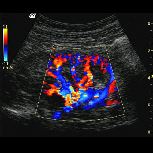
Color Doppler
Doppler Ultrasonography is medical ultrasonography that employs the Doppler effect to generate imaging of the movement of tissues and body fluids(usually blood),and their relative velocity to the probe. By calculating the frequency shift of a particular sample volume, for example flow in an artery or a jet of blood flow over a heart valve, its speed and direction can be determined and visualized. Color Doppler or color flow Doppler is the presentation of the velocity by color scale. Color Doppler images are generally combined with grayscale (B-mode) images to display duplex ultrasonography images, allowing for simultaneous visualization of the anatomy of the area.This is particularly useful in cardiovascular studies (sonography of the vascular system and heart) and essential in many areas such as determining reverse blood flow in the liver vasculature in portal hypertension.
Color Doppler is emerging as a valuable diagnostic imaging modality in the field of reproductive medicine, primarily infertility. Reproductive disorders, including infertility and spontaneousabortions/miscarriages, have emerged as major public health total integral of energy frequency spectrum.Color Doppler is a high-throughput, sophisticated imaging technique for the assessment of uterine anomalies, intrauterine pathology, tubal patency, polycystic ovaries, ovarian follicular monitoring, endometrial receptivity, failed and/or ectopic pregnancy, male infertility, and uterine, endometrial, and ovarian vascularity. Assessments of the uteroovarian pulsatility indices (PIs),resistance indices (RIs), and endometrial color signals are important determinants of in vitro fertilization (IVF) cycles and pregnancy rates. With our clinical/scientific research experience in the field of reproductive medicine, we strongly believe that an overall public health model needs to be designed in managing infertility patients; therefore, major issues such as cost-effectiveness and technical artifacts should be addressed so as to achieve an accurate clinical diagnosis, successful IVF outcome/pregnancy, and overall patient satisfaction in a clinical research setting. Simple, safe, efficient, and affordable diagnostic modalities should be incorporated at infertility clinics coupled with well-designed patient counseling sessions and community-based public health awareness campaigns conducted so as to reduce the morbidity and mortality rates associated with reproductive disorders.